Voluntary movements involve the coordinated activation of two brain pathways that connect parts of deep brain structures called the basal ganglia, according to a study in mice by researchers at the National Institute on Alcohol Abuse and Alcoholism (NIAAA), part of the National Institutes of Health. The findings, which challenge the classical view of basal ganglia function, were published online in Nature on Jan. 23.
“By improving our understanding of how the basal ganglia control movements, these findings could aid in the development of treatments for disorders in which these circuits are disrupted, such as Parkinson’s disease, Huntington’s disease and addiction,” says NIAAA Acting Director Kenneth R. Warren, Ph.D.
The predominant model of basal ganglia function proposes that direct and indirect pathways originating in a brain region called the striatum have opposing effects on movement. Activity of neurons in the direct pathway is thought to promote movement, while activity in the indirect pathway is thought to inhibit movement. Newer models, however, suggest that co-activation of these pathways is necessary to synchronize basal ganglia circuits during movement.
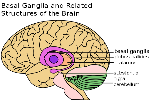
Structure of the basal gandlia, including thalamus, globus paladus, substantia nigra, and cerebellum are shown in this brain image.
“Testing these models has been difficult due to the lack of methods to measure specific neurons in the direct and indirect pathways in freely moving animals,” explains first author Guohong Cui, Ph.D., of the NIAAA Laboratory for Integrated Neuroscience (LIN).
To overcome these difficulties, Dr. Cui and senior author Rui Costa, Ph.D., D.V.M, worked with LIN chief David M. Lovinger Ph.D., NIAAA Cellular Biophotonics Section Acting Chief Steven Vogel, Ph.D., and their colleagues to devise a new approach for measuring the activity of neurons deep within the brain during complex behaviors. Their technique uses fiber optic probes implanted in the mouse brain striatum to measure light emissions from neurons engineered to glow when activated.
Full article at http://neurosciencenews.com/nih-study-advances-understanding-of-movement-control/